Mobile Imaging
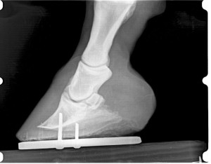 Dr. Seamans relies on state-of-the-art equipment for lameness diagnosis. Mobile digital x-ray and ultrasound are a vital part of modern equine medicine. While conventional radiology (x-ray) allows us to look at bones and structures around joints, ultrasound helps us visualize soft tissues: tendons and ligaments, as well as the cartilage on joint surfaces.
The combination of radiology and ultrasound will locate the problem areas in most cases, and this is necessary before the treatment plan can implemented.
Ultrasound is very useful for reproductive evaluations. Some problems in mares are easily detected with ultrasound. This knowledge can avoid costly delays later in the season. Observation and teasing programs can be useful in managing broodmares. However, early detection of pregnancy with ultrasound is a cheap and reliable way to take the guess work out of monitoring mares.
Pregnancy can be detected as early as 14 days after breeding. This timing is important because twin pregnancies can be more easily managed early. In addition, the absence of a detectable pregnancy by day 14 allows us time to correct some problems without losing a cycle.
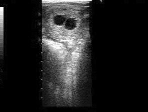
Twin pregnancies usually end in disaster. Early diagnosis with ultrasound makes these relatively easy to manage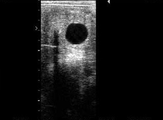
A single pregnancy can be detected at 14 days post breeding.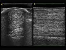
Tendon damage can be seen with ultrasound, sometimes even before swelling or lameness is noticed.
Contact me for your horse vet needs. I offer emergency mobile services.
